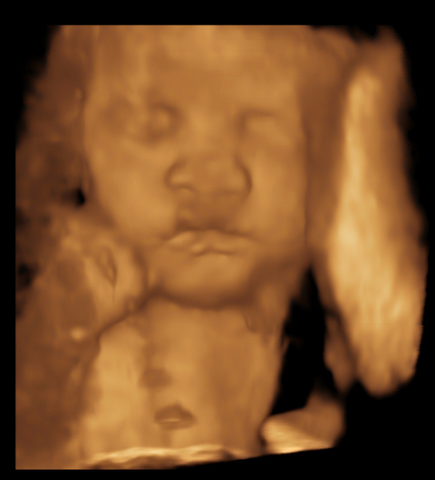
What is 3D and 4D Ultrasound?
In general, ultrasound technology is relatively simple. A machine produces sound waves that are focused on a mother’s uterus. These waves bounce back to the machine and the resulting information forms an image. In the past, the image was similar to a grainy photograph. Three-dimensional ultrasounds, in contrast, actually form a picture that you can manipulate and view every side of the child on a computer screen. The 4D ultrasound incorporates movement within the image, so parents have their first video of the child. Although this technology is known mostly for its use on pregnant women, doctors can use it on other body areas too. A patient with intestinal issues, for example, might have an ultrasound to determine if there’s tears or knots within the digestive tract.
Defining Birth Defects Early On
Both doctors and parents need to know if a child is developing with birth defects. From Down’s Syndrome to cleft palate, some defects can be serious ailments that remain with a child throughout his or her life. Specialized fetal ultrasounds make it possible for doctors to see defects clearly and early on in the pregnancy. Families can plan accordingly when a defect is found. In severe cases, it might be necessary to terminate the pregnancy if the fetus isn’t properly developing and the mother’s life is in danger.
Reflecting Mom and Baby’s Overall Health
When a 3D or 4D ultrasound is performed by a qualified sonogram company, it’s even possible to see the baby’s body in great detail to determine overall health. Pregnant women usually take prenatal pills to enhance their mineral and vitamin needs as the baby consumes much of these nutrients. If the mother isn’t leading a healthy lifestyle, any malnutrition issues will reflect in the baby’s detailed ultrasound. Doctors might see limbs developing with skinny shapes, indicating that calcium deficiencies could be present. With detailed nutrient information on-hand, doctors advise their patients on alternative diets to keep the baby as healthy as possible.
Evaluating Baby’s Organ Development
Modern ultrasounds also make it possible for doctors to monitor the fetus’s internal organs. They can clearly examine the heart’s activity and verify that no irregular rhythms are present, for instance. The brain’s developing folds are another area that doctors examine very closely. Although ultrasounds shouldn’t be performed on a constant basis, one or two appointments can give doctors enough information to accurately care for the mom and baby when everything looks normal. As more doctors use this technology, they’ll have a better insight into the developing fetus than ever before.
When you decide to have a 3D or 4D ultrasound, it’s important to select the right facility for your procedure. An accredited sonogram professional must be on-hand to perform and decipher the images. Your first baby picture will be an amazing one with experienced professionals capturing the image.
